New research has found a new type of can could be used to assess the effectiveness of chemotherapy given before surgery.
New research funded by Breast Cancer Now has found a new type of ultrasound scan could be used to accurately assess the effectiveness of chemotherapy given before surgery.
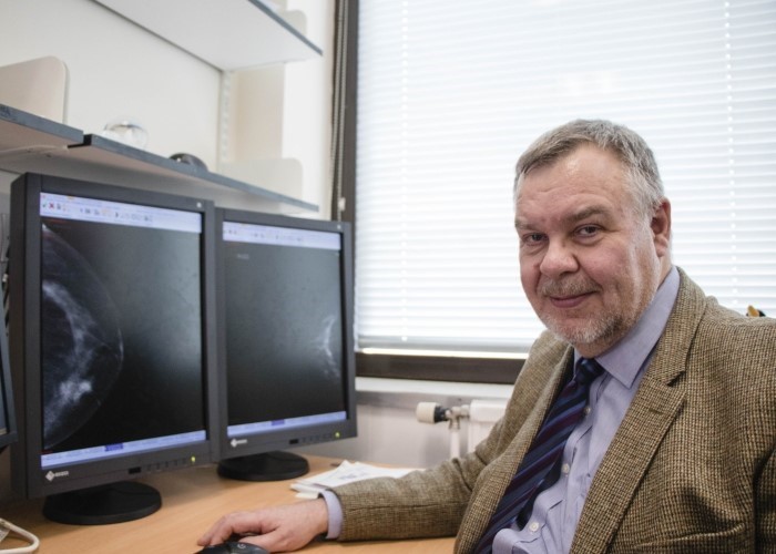
Photo credit: University of Dundee
Chemotherapy is often used as a treatment for breast cancer and, thanks to new research, a new type of scan could be used to assess the effectiveness of it given before surgery.
New findings
Research funded by Breast Cancer Now has found a new type of ultrasound scan could be used to accurately assess the effectiveness of chemotherapy given before surgery.
Scientists at the University of Dundee found the new ultrasound works as well as an MRI scan to recognise whether breast cancer cells remain at the site of a breast tumour after chemotherapy.
Researchers hope this new method could provide a faster, cheaper and more widely available way to decide the best course of treatment for people with breast cancer.
It could also help some patients avoid more extensive surgery.
The impact of surgery
Chemotherapy is given before surgery to shrink a larger tumour in the breast or to slow down its growth if it’s growing rapidly. If the tumour shrinks, it sometimes means patients can have breast-conserving surgery rather than a mastectomy. If chemotherapy clears all signs of cancer from the breast, it could also mean a smaller number of lymph nodes have to be removed by surgery.
Dr Kotryna Temcinaite, Research Communications Manager at Breast Cancer Now, explained the impact that breast cancer surgery can have on patients.
‘We know that having breast cancer surgery can have a significant emotional and physical impact on many patients and there are risks and side effects to surgery. This is a really promising finding that could in future help some patients avoid more extensive breast cancer surgery.’
In this new study, researchers led by Professor Andrew Evans at the University of Dundee worked with 80 breast cancer patients who were due to receive chemotherapy before surgery. The researchers tested whether breast cancer cells were still present at the time of surgery and if so whether they were detected by different imaging methods.
A new wave technology
A standard ultrasound scan uses sound waves to produce an image of the breast tissue.
Shear wave elastography is an exciting new technology that scientists believe could be added to standard ultrasound to improve how we image breast cancer tumours. The technique relies on measuring a tissue’s ‘stiffness’, and means we can tell apart scar tissue left after chemotherapy from a remaining breast cancer tumour.
Professor Andrew Evans explained the importance of these findings.
‘Our study is the first time that a comparison of this nature has been undertaken and we found that shear wave elastography combined with ultrasound had a similar accuracy to MRI. It’s quick and reproducible so it could potentially replace MRI in the future, which is expensive, time-consuming and cannot be given to some women.
‘This is an important finding that could have a real impact on patients’ treatment in the future, and we now need to see larger studies to verify our findings.’
Kotryna added, ‘As more effective treatments become available, this new method could even help some patients to avoid having surgery altogether, which could have a major impact on their quality of life.’
The researchers suggest that shear wave elastography could be a way to assess people who can’t have an MRI, due to metal objects in their body or implants such as pacemakers.
Quicker and cheaper
Currently, standard ultrasound or MRI is used to assess how a tumour has responded to chemotherapy given before surgery. Greyscale ultrasound is a widely used technique that is quick and cheap. However, it can’t always to tell apart leftover cancer cells form scar tissue after chemotherapy.
Shear wave elastography, combined with an ultrasound scan, is faster than MRI. The new technique takes around 4 minutes to give results compared to 30–45 minutes for MRI. The technique is also cheaper, costing around £200 less than a breast MRI.
‘New imaging methods like this will be vital in helping clinicians and patients decide on the most effective course of treatment, which in turn will help achieve the best possible quality of life after treatment for breast cancer patients,’ said Kotryna.
This research was published in Clinical Radiology.
If you have any concerns about breast cancer surgery or treatment, you can speak to our expert nurses on our free Helpline at 0808 800 6000 or by using our confidential Ask Our Nurses service.